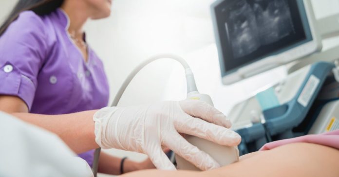
What is Ultrasound examination?
Ultrasonography is a diagnostic imaging technique that involves scanning the body’s internal organs using high-frequency sound waves. The image shows the size, density, and shape of internal organs to help in medical diagnosis.
What is ultrasound used for?
Diagnostics
Physicians use ultrasound imaging to help diagnose conditions affecting the organs and soft tissues of the body, including:
- Liver
- Abdomen
- Heart
- Spleen
- Kidneys
- Blood vessels
- Gallbladder
- Thyroid
- Pancreas
- Breast
- Bladder
- Ovaries
- Eyes
- Testicles
There are certain diagnostic limitations for ultrasounds. For instance, sound waves do not transmit well through areas that may hold gas or air (like intestines), or areas blocked by dense bone.
Therapeutic application
Sometimes, ultrasound is used in the detection and treatment of certain soft-tissue injuries.
Medical procedures
When a physician has to remove tissue from a very precise area in your body (such as in a needle biopsy) ultrasound imaging can help by giving visual direction.
What is the role of USG?
Ultrasound scans are usually associated with pregnancy. The scans can give a mother anticipating delivery the first view of her unborn child. The test is not limited only to pregnancy and helps diagnose medical conditions such as kidney stones, gallstones, enlarged organs, and liver disease.
A doctor can ask for an ultrasound scan if a person is suffering pain, swelling, or displays other symptoms that require an internal view of the organs. Ultrasound sonography is used for examination of the internal organs without any incisions. Some of the internal organs seen by ultrasonography include:
- Bladder
- Brain (in infants)
- Eyes
- Gallbladder
- Liver
- Kidneys
- Pancreas
- Ovaries
- Spleen
- Thyroid
- Testicles
- Uterus
- Blood vessels
Ultrasonography is also used to guide surgeons’ movements during some medical procedures like biopsies.
How to prepare for an ultrasound?
The preparation for ultrasound sonography depends on the area of the body or the organ being examined. Different preparations depending on the organs are:
- The doctor might advise the patient to fast for 8 to 12 hours before the abdomen ultrasound is conducted as undigested food can block the sound waves, thus hindering the process of getting a clear image.
- The examination of the gallbladder, liver, pancreas, or spleen might require consuming a fat-free meal and fast till the procedure. They can, however, continue to have water and take any medications prescribed by the doctor.
- A person might be asked to drink a lot of water for viewing the pelvic organs so that the bladder is full and visualized better.
How is an ultrasound performed?
Ultrasound Sonography is performed as follows:
- The patient changes into a hospital gown and may be advised to lie down on the examination table with a section of the body exposed for the test.
- A special lubricating jelly will be applied to the skin by an ultrasound technician called a sonographer to prevent friction and rub the ultrasound transducer on the skin. The jelly helps in the transmission of the sound waves.
- High-frequency sound waves sent through the body by the transducer, which reverberate as they hit a dense object like a bone or an organ. The reverberations are then reflected into a computer.
- The sound waves form a picture that the doctor can interpret
- A person might need to change positions depending on the area being examined so the technician can have better access.
- The gel will be cleaned off of the skin after completing the procedure. It typically lasts less than 30 minutes and depends on the area being examined.
- As soon as the procedure is completed, the patient will be free to go about with their normal activities.
What happens after an ultrasound?
Following an ultrasound, the doctor will check and review the images for any abnormalities.
The patient will be called, or a follow-up will be scheduled for the discussions regarding the findings from the examination. The patient may have to undergo other diagnostic techniques like MRI, CT scan, or a biopsy sample of tissue depending on the area examined if anything abnormal turns up on the ultrasound.


